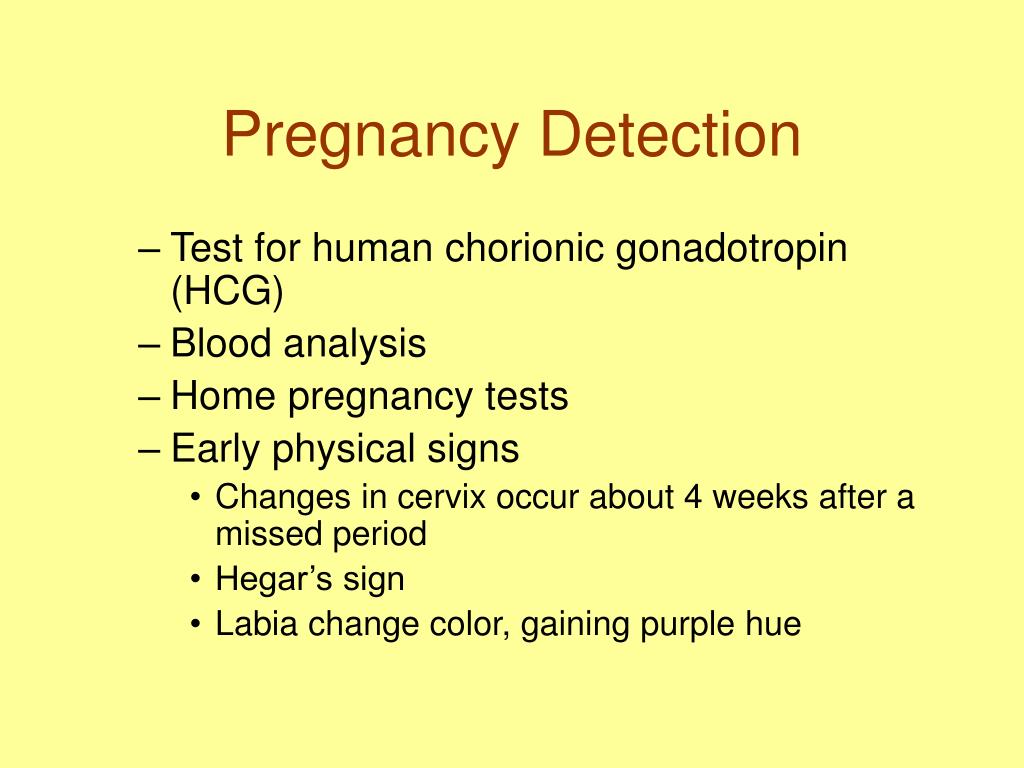

The beta subunit determines the unique physiological, biochemical, and immunological properties of hCG. The alpha subunit is identical to the alpha subunits of luteinizing hormone (LH), follicle-stimulating hormone (FSH), and thyrotropin (TSH, formerly thyroid-stimulating hormone), while the beta subunit has significant homology to the beta subunit of LH and limited similarity to the FSH and TSH beta subunits. HCG is a glycoprotein consisting of 2 noncovalently-bound subunits. Low levels of estriol also have been associated with pregnancy loss, Smith-Lemli-Opitz, and X-linked ichthyosis (placental sulfatase deficiency). Decreased uE3 has been shown to be a marker for Down syndrome and trisomy 18. Estriol levels increase during the course of pregnancy. The half-life of uE3 in the maternal blood system is 20 to 30 minutes because the maternal liver quickly conjugates estriol to make it more water soluble for urinary excretion. Estriol exists in maternal blood as a mixture of the unconjugated form and a number of conjugates. Lower maternal serum AFP values have been associated with an increased risk for genetic conditions such as Down syndrome and trisomy 18.Įstriol, the principal circulatory estrogen hormone in the blood during pregnancy, is synthesized by the intact feto-placental unit.

Additionally, increased maternal serum AFP values may be seen in pregnancies with multiple fetuses and in unaffected singleton pregnancies in which the gestational age has been underestimated. Other fetal abnormalities such as omphalocele, gastroschisis, congenital renal disease, esophageal atresia, and other fetal-distress situations such as threatened abortion and fetal demise, may also show AFP elevations.

Subsequently, the AFP reaches the maternal circulation, thus producing elevated serum levels. If the fetus has an open NTD, AFP is thought to leak directly into the amniotic fluid causing unexpectedly high concentrations of AFP. The AFP concentration in maternal serum rises throughout pregnancy, from a nonpregnancy level of 0.2 to about 250 ng/mL at 32 weeks gestation.

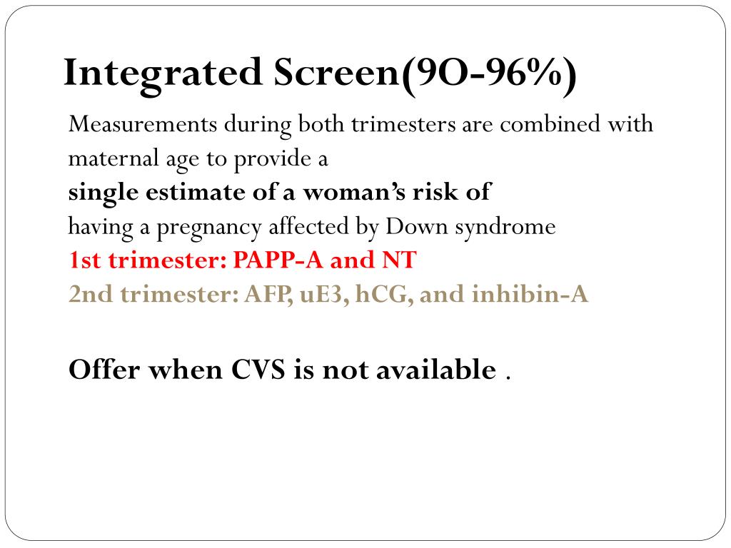
A small amount also is transported from the amniotic cavity. Fetal AFP diffuses across the placental barrier into the maternal circulation. The concentration of AFP peaks in fetal serum between 10 to 13 weeks. By the end of the first trimester, nearly all of the AFP is produced by the fetal liver. A small amount also is produced by the gastrointestinal tract. If results are positive, the patient is typically offered counseling and diagnostic testing.ĪFP is a fetal protein that is initially produced in the fetal yolk sac and liver. The information from both trimesters is combined and a report is issued. The blood sample is tested for AFP, unconjugated estriol (uE3), human chorionic gonadotropin (hCG), and inhibin A. If the risk from the first trimester is below the established cutoff, an additional serum specimen is collected in the second trimester for this test. For a stand-alone NTD-risk assessment, order MAFP1 / Alpha-Fetoprotein (AFP), Single Marker Screen, Maternal, Serum. When the part 1 screen is completed, a NTD risk is not provided. In that event, the patient is typically offered counseling and diagnostic testing. If the result from part 1 indicates a risk for Down syndrome that is higher than the screen cutoff, the screen is completed, and a report is issued. The results of the ultrasound measurement and blood work along with the maternal age and demographic information are used to calculate Down syndrome and trisomy 18 risk estimates. Along with the NT measurement, a maternal serum specimen is collected to measure pregnancy-associated plasma protein A. Therefore, NT data is accepted only from NT-certified sonographers. The ultrasound measurement, referred to as the NT measurement, is difficult to perform accurately. SEQA / Sequential Maternal Screening, Part 1, Serum involves an ultrasound and a blood collection. Sequential screening combines biochemical and ultrasound markers (nuchal translucency: NT) measured in both trimesters of the pregnancy. Sequential screening is a type of cross-trimester screening, which has an improved detection rate as compared to either first- or second-trimester screening. Various options for maternal serum screening are available and include: first trimester, second trimester, and cross-trimester. Maternal serum screening is used to identify pregnancies that may have an increased risk for certain birth defects, such as trisomy 21 (Down syndrome), neural tube defect (NTD) and trisomy 18.


 0 kommentar(er)
